


Plaster cast
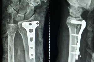
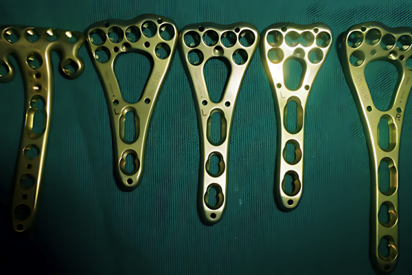
Plating

Kirschner wires


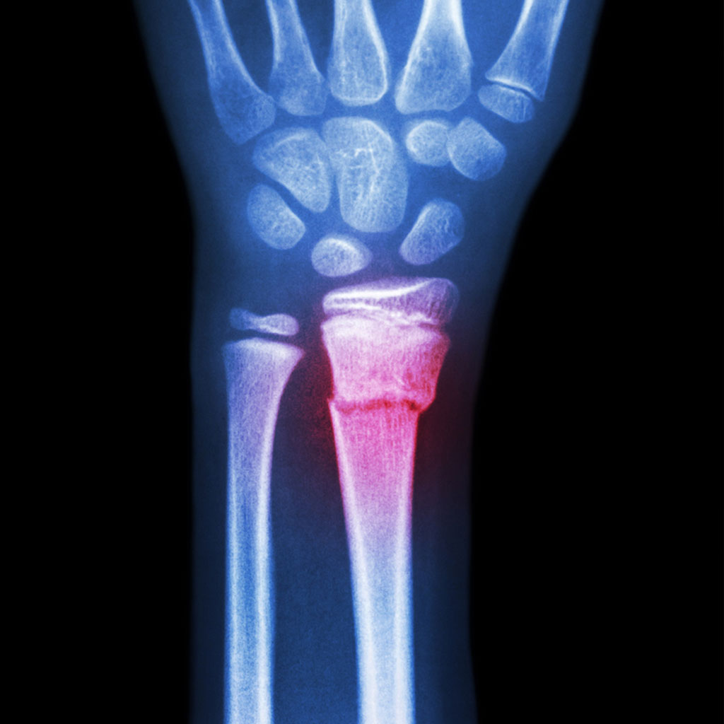
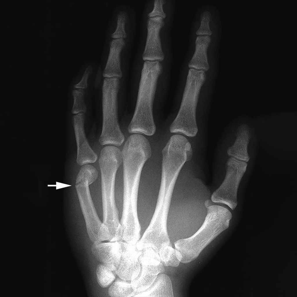
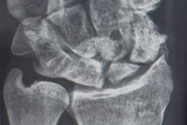
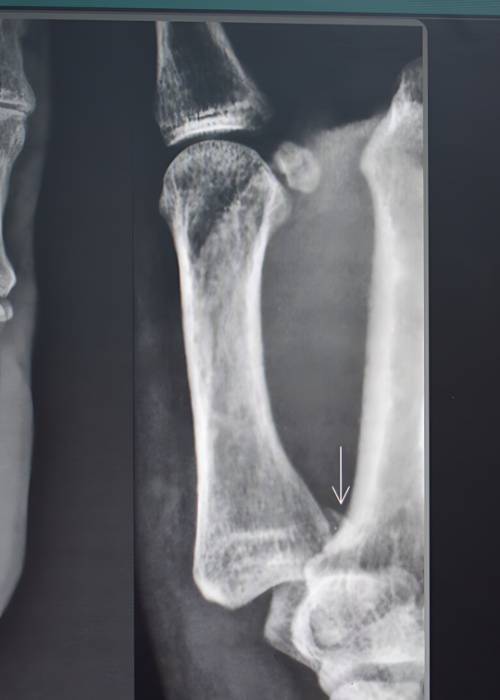
Diagnosis
An x-ray reveals, loss of alignment of the bones. Interpreting the x-ray properly helps to avoid missing the diagnosis. At times, the report accompanying the x-ray film may be erroneous, misleading and delays establishing the correct diagnosis. A good quality x-ray image with the appropriate position of the finger and magnification will show loss of collinear phalanges. Once the loss of alignment is established, the next step, is to assess the extent of joint affection and whether the fracture has resulted in gross multiple pieces (comminution).


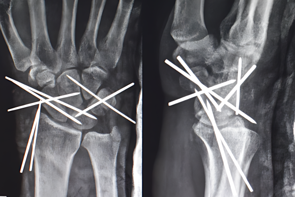
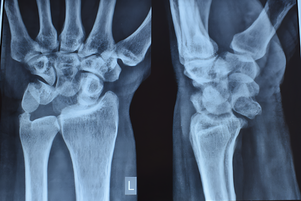

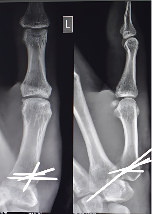


After Surgery
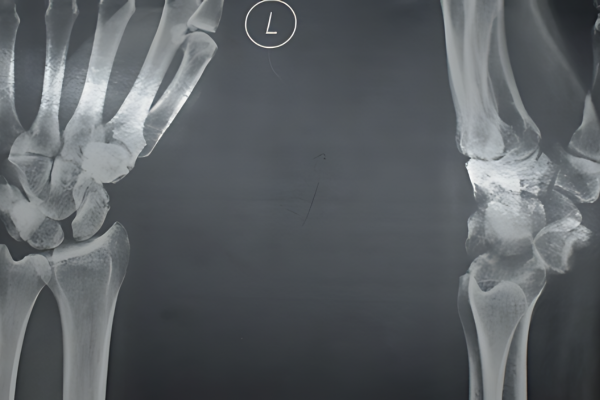
Before Surgery


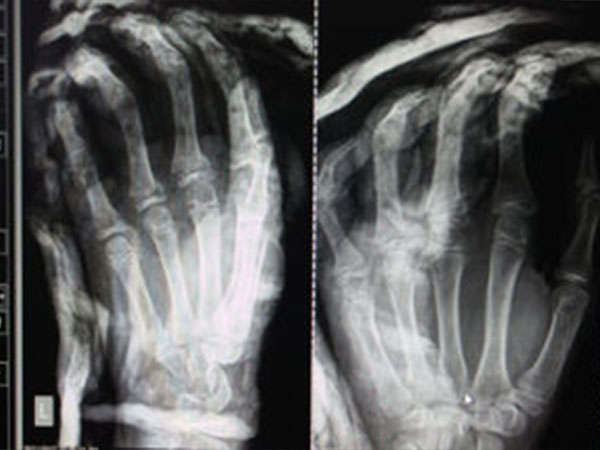
After Surgery
Before Surgery